Research Themes:
Our research program is based on targeted pathology phenotyping, which uses the morphology of early disease lesions as the “disease phenotype” rather than global spirometry measures. The program uses a multi-level approach ranging from clinical CT imaging of patient cohorts, biobanking of human tissues and cells, single cell resolution technologies and in vitro disease models.
1. Understanding the contribution of dysregulated epithelial repair to airway remodelling in asthma:
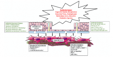
The epithelial-mesenchymal trophic unit (EMTU) involves communication between the structural cells of the lung (epithelium and mesenchyme), inflammatory cells, and their environment (extracellular matrix). The EMTU in the healthy lung regulates key processes including lung development and repair, but in disease dysregulation of this cross-talk leads to inflammation and tissue remodeling. We have developed and phenotyped several multi-cellular lung disease models to understand the contribution of structural cells in the EMTU to inflammation and remodeling in asthma and COPD.
References:
Epithelial-mesenchymal crosstalk in COPD: An update from in vitro model studies – ScienceDirect
2. Development of microCT imaging methodology to understand small airways disease:
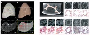
The Central Hypothesis of our research program is that disruption of normal repair processes within the EMTU of the lung propagates inflammation and remodeling leading to disease. The goal of our research program is to move from clinical spirometry measures of whole organ lung function that have, in the past, provided weak associations for therapeutic testing. Instead, we use novel micro x-ray Computed Tomography (micro CT) imaging to identify disease lesions and then conduct molecular phenotyping studies to identify the true molecular signatures of each airway disease. To this end my group, in collaboration, has developed several novel imaging protocols using micro CT to image samples frozen or embedded in paraffin.
References:
https://www.thelancet.com/journals/lanres/article/PIIS2213-2600(18)30196-6/fulltext
3. Development of imaging methodology to understand multi-cellular tissues:
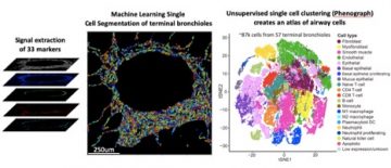
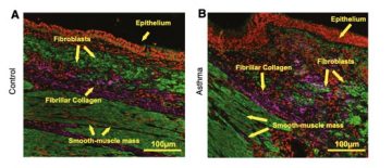
To understand the focal nature of pathology in chronic diseases, our lab developed several imaging methodologies to quantify the 3-dimentional (3D) interactions of cells and the extra cellular matrix (ECM) using confocal, non-linear optical microscopy (NLOM) and imaging mass spectrometry. These methodologies have been used with many of the 3D tissue culture systems we have developed in my laboratory to understand epithelial cells and fibroblast interactions within the lung EMTU. We have demonstrated how NLOM, compared to traditional histological techniques, can be harnessed to understand how structural alterations in collagen and elastin fibers can contribute to airway remodeling in asthma and COPD. Most recently, we have developed imaging CyTOF mass spectrometry to analyze whole mount tissue sections with up to 35 antibodies, to understand the complex tissue environment in disease pathologies.
References: